At the scene of a crime
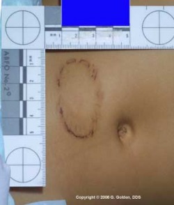
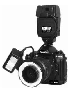
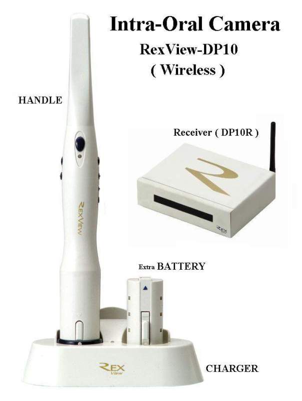
Upon finding bitemarks it is important to document it. That means recording the injury, narrative notes on its physical appearance, colour, size and where it is on the body. It is important to take note of the contour and elasticity of its location on the body. When taking photographs it is necessary to use both a extraoral and intraoral camer equiped with macro lens and both black-and-white and colour film. The camera should be positioned directly over the bitemark are so that the area isn't distorted. After photographing the injury the next step is too take a saliva swab of the area to try and gather any of DNA evidence. The two swab technique is used when gathering this type of evidence: first cotton swab is moistened with distilled water and used to wash the surface of the bite area with circular motions and applying light pressure. The second swab is with a dry one that collects the moisture on the bitemark. Both swabs are then air-dried for fourty-five minutes before being released to the authorites for testing.
Another method for extracting DNA is the "Cryogenic Grinding of Teeth and Bones" technique. Complete guide for this technique can be found on BOLD's website on this link. http://www.boldlab.org/pdf/cryo.grind.pdf
Another method for extracting DNA is the "Cryogenic Grinding of Teeth and Bones" technique. Complete guide for this technique can be found on BOLD's website on this link. http://www.boldlab.org/pdf/cryo.grind.pdf
Impressions
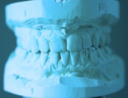
Dental impressions are an accurate representation of person's dentition. The dental impressions of teeth and gums form a "negative" which is then transfigured into a cast or model of the dentition. Materials generally used for creating an impression are: Sodium alginate, polyether and silicones (condensation and addition-cured silicones ex. polyvinyl siloxan). After the impressions are created a plaster is poured into creating a plaster model, these model's are then scanned to make a digital image. A computer program called Lucis is used to enhance the patterns in the bite mark, which are then traced and superimposed onto a picture of the bite mark. Before the program Lucis was created it was very hard toget a good quality photograph of the bitemarks as the body's healing process starts to take away the bitemark's characteristics very quickly. However if the bitemark's were after the death then they wouldn't diminish because the healing process cease's upon death.
Bitemark analysis studies are done using pig skin because it is comparable with human skin. However there are many limitations from this because while the two are comparable there are still many differences. For example stimulated pressures are used to create bitemarks; however pig and human skin have different elasticity. Also the postmortem bites on a nonhuman skin display different bite patterns compared to those in antemortem bite patterns.
Other methods of bite mark analysis was using the "fingerprint powder lift-models". With this technique the bitten skin would be dusted with black fingerprint powder and then lifted onto a sheet of acetate. However more recent developments are such as the Lucis software program.
